Diagram Of Shoulder / Anatomy Of The Rtc Tendons Right Shoulder Download Scientific Diagram. Numerous muscles help stabilize the three joints of. Basic shoulder anatomy the shoulder complex is made up of three bones, which are connected by muscles, ligaments, and tendons. There are two joints within the shoulder that can be affected by osteoarthritis. The following is an overview of the shoulder muscle anatomy. The scapula shoulder blade is a triangular shaped bone with 2 bony projections at the top right at your shoulder cuff.
What are common rotator cuff injuries? These muscles form the outer shape of the shoulder and underarm. A metal ball component replaces the worn humeral head. The supraspinatus is located on the upper part of the shoulder joint and is involved in abduction (arm raising). Is the wear and tear of shoulder cartilage until bare bone is exposed.
Exam Series Guide To The Shoulder Exam Canadiem from canadiem.org Hold each position for a total of five breaths, while attempting to strongly contract the muscles that create each distinct motion. A dislocated shoulder occurs when the humerus (upper arm bone) separates from the shoulder blade at the main shoulder joint. The shoulder joint (glenohumeral joint) is a ball and socket joint between the scapula and the humerus.it is the major joint connecting the upper limb to the trunk. Shoulder pain that persists beyond a few days. The shoulder blade is called the scapula and the collarbone is called the clavicle. The most flexible joint in the entire human body, our shoulder joint is formed by the union of the humerus, the scapula (or shoulder blade), and the clavicle (or collarbone). The shoulder girdle includes three bones—the scapula, clavicle and humerus. Learn about these muscles, their origin and insertion points, and their functional anatomy.
The treatment options are either replacement of just the head of the humerus bone (ball), or replacement of both the ball and the socket (glenoid).
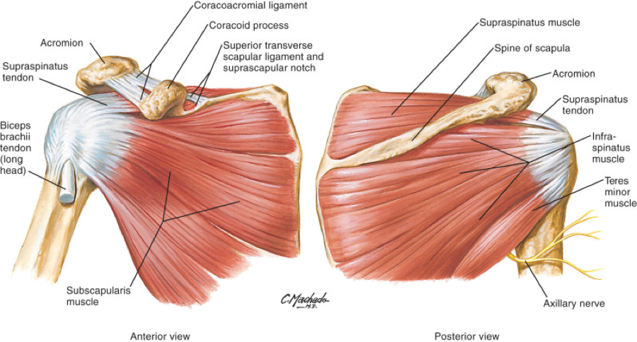
The following shoulder range of motion diagram contains images of 12 different humerus, scapula and clavicle moves. It is one of the most mobile joints in the human body, at the cost of joint stability. The main shoulder muscles are trapezius, deltoid, pectoralis major and 4 rotator cuff muscles: Subscapularis, supraspinatus, infraspinatus and teres minor. Basic shoulder anatomy the shoulder complex is made up of three bones, which are connected by muscles, ligaments, and tendons. Numerous muscles help stabilize the three joints of. The shoulder girdle includes three bones—the scapula, clavicle and humerus. A metal ball component replaces the worn humeral head. Bones in shoulder, ligaments of the shoulder joint, parts of the shoulder joint, shoulder anatomy, shoulder joints and muscles, shoulder structure anatomy, shoulder tendon anatomy, shoulder tendons ligaments, human muscles, bones in shoulder, ligaments of the shoulder joint, parts of. The acromioclavicular joint is where the acromion, part of the shoulder blade (scapula) and the collar bone (clavicle) meet. The shoulder joint is composed of the glenoid (the shallow shoulder socket) and the head of the upper arm bone known as the humerus (the ball). Pain in a man's body pain in a man's body on a gray background. The shoulder joint is formed where the humerus (upper arm bone) fits into the scapula (shoulder blade), like a ball and.
This is the main muscle that lets you rotate and extend your shoulder. The treatment options are either replacement of just the head of the humerus bone (ball), or replacement of both the ball and the socket (glenoid). Inside the shoulder there are three joints; The glenohumeral joint is a joint where the greater tubercle (humeral head at the top of the arm bone) meets the shoulder socket of the scapula, called the glenoid cavity or glenoid fossa. If you have new, worsening, or severe shoulder pain, you should seek medical attention.
Labeled Anatomy Chart Of Neck And Shoulder Muscles On Black Background Stock Photo Download Image Now Istock from media.istockphoto.com Shoulder pain that persists beyond a few days. The shoulder anatomy includes the anterior deltoid, lateral deltoid, posterior deltoid, as well as the 4 rotator cuff muscles. The muscles in the shoulder aid in a wide. The glenohumeral joint is where the ball (humeral head) and the socket (the glenoid) meet. Subscapularis, supraspinatus, infraspinatus and teres minor. The shoulder joint (glenohumeral joint) is a ball and socket joint between the scapula and the humerus.it is the major joint connecting the upper limb to the trunk. The large bone in the upper arm is called the humerus. Smartdraw includes 1000s of professional healthcare and anatomy chart templates that you can modify and make your own.
A second joint in the shoulder is the junction of the collar bone with the shoulder blade, called the.
On the left is a standard (anatomic) shoulder arthroplasty. Diagram of the shoulder, including the location of the rotator cuff. It is one of the most mobile joints in the human body, at the cost of joint stability. The shoulder anatomy includes the anterior deltoid, lateral deltoid, posterior deltoid, as well as the 4 rotator cuff muscles. The main shoulder joint, called the glenohumeral joint, is formed The main shoulder muscles are trapezius, deltoid, pectoralis major and 4 rotator cuff muscles: This is the smallest rotator cuff muscle. The supraspinatus is located on the upper part of the shoulder joint and is involved in abduction (arm raising). Basic shoulder anatomy the shoulder complex is made up of three bones, which are connected by muscles, ligaments, and tendons. Inability to carry objects or use your arm. The primary function of the shoulder girdle is to give strength and range of motion to the arm. The treatment options are either replacement of just the head of the humerus bone (ball), or replacement of both the ball and the socket (glenoid). Its main job is to assist with rotation of the arm away from the body.
Your mission is to try them out on your body. The roof of the shoulder is formedby a part of the scapula called the acromion. The list of muscles and their functions are presented below. Is the wear and tear of shoulder cartilage until bare bone is exposed. Inside the shoulder there are three joints;
Anatomy Of Shoulder Joint And Shoulder Girdle Download Scientific Diagram from www.researchgate.net The main shoulder muscles are trapezius, deltoid, pectoralis major and 4 rotator cuff muscles: A dislocated shoulder occurs when the humerus (upper arm bone) separates from the shoulder blade at the main shoulder joint. The large bone in the upper arm is called the humerus. The shoulder is a complex combination of bones and joints where many muscles act to provide the widest range of motion of any part of the body. The humeral head is the ball side. The shoulder joint can sometimes become narrowed and arthritic, and spurs can form on the undersurface. It is one of the most mobile joints in the human body, at the cost of joint stability. The shoulder joint is the junction between the chest and the upper extremity.
Pronate your wrist so the palm of your hand faces down to the floor (as if you were trying to empty a glass of water).
The primary function of the shoulder girdle is to give strength and range of motion to the arm. While seated or standing, lift the sore arm forward and to the side about thirty to 45 degrees. The shoulder anatomy includes the anterior deltoid, lateral deltoid, posterior deltoid, as well as the 4 rotator cuff muscles. Shoulder pain that persists beyond a few days. The main shoulder joint, called the glenohumeral joint, is formed The components of the ball and cup are reversed on the right—a reverse shoulder replacement. The scapula shoulder blade is a triangular shaped bone with 2 bony projections at the top right at your shoulder cuff. What are common rotator cuff injuries? The glenohumeral joint is where the ball (humeral head) and the socket (the glenoid) meet. The acromioclavicular joint is where the acromion, part of the shoulder blade (scapula) and the collar bone (clavicle) meet. Plus, exercises for training them. Numerous muscles help stabilize the three joints of. This is the smallest rotator cuff muscle.
Diagram Of Shoulder / Anatomy Of The Rtc Tendons Right Shoulder Download Scientific Diagram. There are any Diagram Of Shoulder / Anatomy Of The Rtc Tendons Right Shoulder Download Scientific Diagram in here.